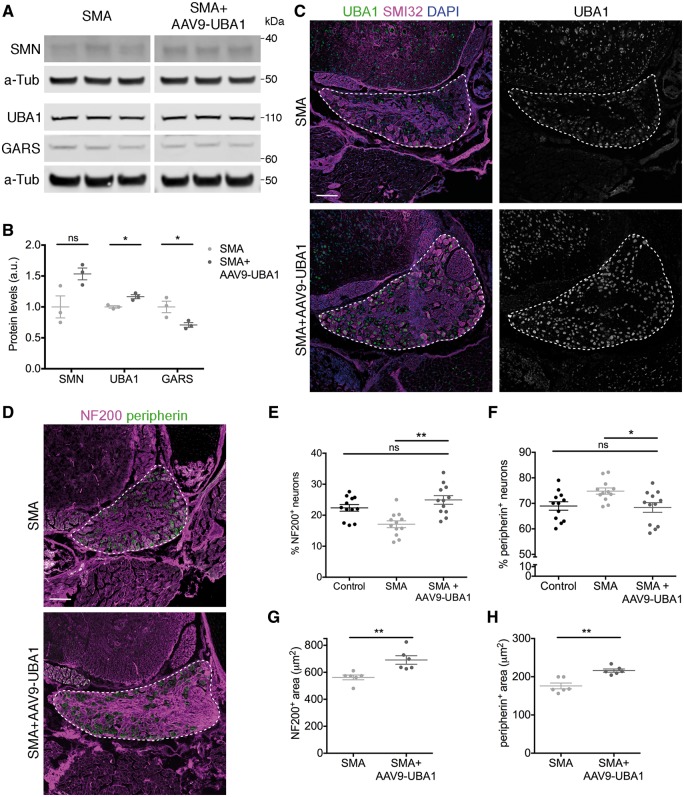
Figure 7.
Restoration of UBA1 in SMA mice reverses GARS dysregulation and rescues sensory neuron cell fate phenotypes. (A and B) Representative fluorescent western blot (A) and quantification (B) of SMN, UBA1 and GARS in dorsal root ganglia from late-symptomatic SMA mice and SMA mice injected with AAV9-UBA1 (SMA+AAV9-UBA1). α-Tubulin (α-Tub): loading control. n = 3 mice per condition. (C) Spinal column sections from lumbar segments 1 and 2 of late-symptomatic SMA and SMA+AAV9-UBA1 mice labelled with UBA1a (green), SMI32 (magenta) and DAPI. (D) Spinal column sections from lumbar segments 1 and 2 of late-symptomatic SMA and SMA+AAV9-UBA1 mice labelled with NF200 (magenta) and peripherin (green). (C and D) Dorsal root ganglia (DRG) are outlined. (E and F) Quantification of the percentage of NF200-positive (NF200+) (E) and peripherin-positive (peripherin+) (F) sensory neurons. Data from control mice is shown as reference. n = 3 mice per condition, n = 4 DRGs per mouse. (G and H) Quantification of the area of NF200+ (G) and peripherin+ (H) sensory neurons. n = 3 mice per condition, n = 2 DRGs per mouse (seven NF200+ and seven peripherin+ neurons per DRG were analysed); ns = not significant. *P < 0.05, **P < 0.01. See also Supplementary Figs 10 and 11.

