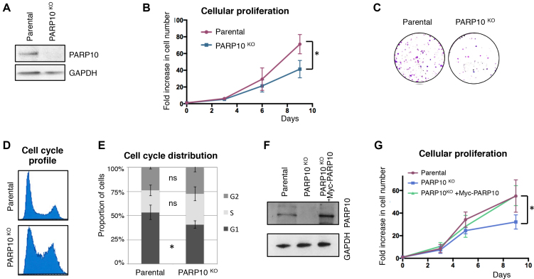
Figure 1.
Loss of PARP10 impairs proliferation of HeLa cells. (A) Western blot showing loss of PARP10 expression in HeLa cells with CRISPR/Cas9-mediated PARP10 knockout. (B) PARP10-knockout HeLa cells show reduced proliferation rates. The average of three experiments with error bars representing standard deviations is shown. The asterisk indicates statistical significance (using the two-tailed equal variance TTEST). (C) Representative clonogenic assay showing reduced proliferation of PARP10-knockout HeLa cells. (D) Representative PI flow cytometry profile showing an altered cell-cycle distribution in PARP10-knockout HeLa cells. (E) Quantification of cell-cycle distribution in control and PARP10-knockout HeLa cells. The average of four experiments, with error bars as standard deviations, is shown. Statistical significance was calculated using the two-tailed equal variance TTEST. (F) Western blot showing the re-expression of PARP10, with a Myc-tag, in PARP10-knockout HeLa cells. (G) Exogenous PARP10 expression rescues the proliferation defect of PARP10-deleted HeLa cells. The average of four experiments with error bars representing standard deviations is shown. The asterisk indicates statistical difference between the PARP10KO and PARP10KO + Myc-PARP10 samples.

