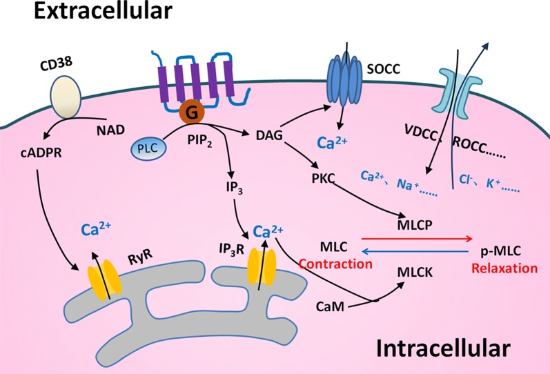
Fig. 2.
Mechanisms of contraction in ASM. Many regulatory mechanisms in ASM to control the contraction and relaxation are well recognized. The ectoenzyme CD38 could produce the second messenger cyclic ADP ribose (cADPR), which causes Ca2+ release through ryanodine receptor (RyR) channels from sarcoplasmic reticulum (SR). The G-protein coupled receptor (GPCR)-based pathway activates phospholipase C (PLC) and breaks up phosphatidyl diphosphate inositol 2 (PIP2) into inositol trisphosphate (IP3) and diacylglycerol (DAG). Intracellular Ca2+ binds to intracellular calmodulin (CAM) to alter the phosphorylation status of myosin light chain (MLC) and regulates ASM function. Depletion of Ca2+ in SR calcium stores induces Ca2+ influx through store-operated Ca2+ channel (SOCC). There are other mechanisms that take part in regulating the intracellular ion concentration of ASM and finally affect the contraction and relaxation of them

