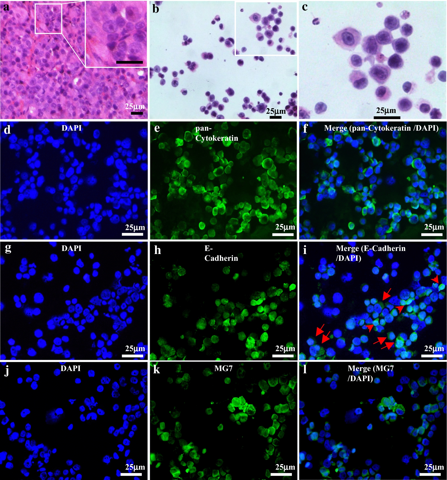
Fig. 2.

Morphologic and immunohistochemical features of cells retrieved from the implanted capsules in MiniPDX-bearing mice. a Tissue section of a PDX xenograft tumor (GASI80) showing typical feature of poorly differentiated adenocarcinoma; inlet: High magnification view revealing tightly arranged poorly differentiated cells. b, c Cytospin of cells retrieved from the capsules implanted in MiniPDX-bearing mice, low- and high-power view, respectively (H&E stain), showing that the majority of the cells are associated with high nucleus to cytoplasm ratio, hyperchromatic nuclei, and scant cytoplasm. Immunofluorescent staining of pan-cytokeratin (e), E-cadherin (h) and MG7 (i); 4′, 6-diamidino-2-phenylindole staining for individual panels (d, g, j). Merged images (f, i, l) show that the cells cultivated within the OncoVee® capsules expressed all of the three characteristic primary gastric cancer-related markers. Scale bars, 25 μm. The tumor cells cultivated in the MiniPDX capsules, which were derived from PDX tumor of gastric adenocarcinomas (PDX model GASI80, a H&E stain of tissue section), strongly expressed pan-cytokeratin (e, f) E-cadherin (h, i), and MG7 (k, l)
