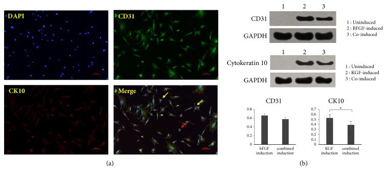
Figure 5.
(a) Immunofluorescence staining of DAPI, CD31, and CK10 of iPSC-MSCs after combined induction (×100). (b) Results of Western blot assay, indicating that bFGF or KGF induction could enhance the expression of CD31 or CK10, whereas in the combined induction group, the expression of CD31 and K10 was lower than that of the single induction group. ∗P<0.05. The red arrow indicated that only a small number of cells showed a tendency towards shifting into a round shape after induction; meanwhile the yellow arrow showed that most of the cells still exhibited a MSC-like long spindle type.

