Figure 1.

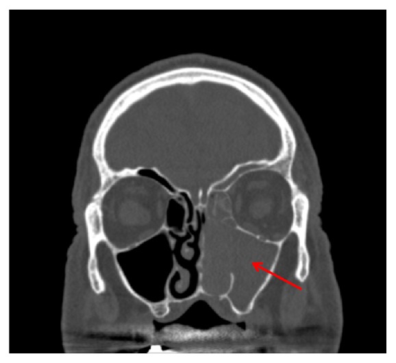
CT sinuses (coronal view) for Case 1. There is contiguous opacification of the left maxillary sinus, nasal cavity, ethmoid air cells, and frontal sinus.

CT sinuses (coronal view) for Case 1. There is contiguous opacification of the left maxillary sinus, nasal cavity, ethmoid air cells, and frontal sinus.