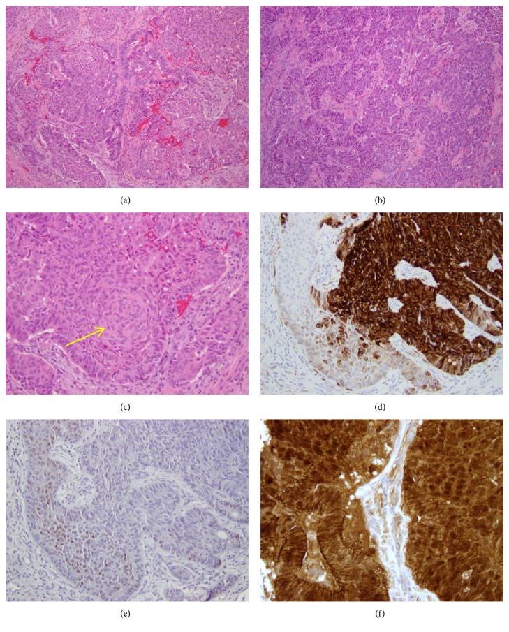
Figure 2.
Morphologic and immunohistochemical findings in Case 1. The tumor is composed of a mixture of back-to-back glands, anastomosing cords, and solid areas with squamoid morular metaplasia ((a) and (b), H&E, x100). Squamoid morular metaplasia is easily found ((c), H&E, x200). CK7 is positive in areas of glandular differentiation and negative in squamoid morules ((d), x200). CDX2 is positive in squamoid morules and negative in glandular areas ((e), x200). β-catenin shows diffuse membranous staining throughout the tumor and nuclear staining restricted to squamoid morules ((f), x400).

