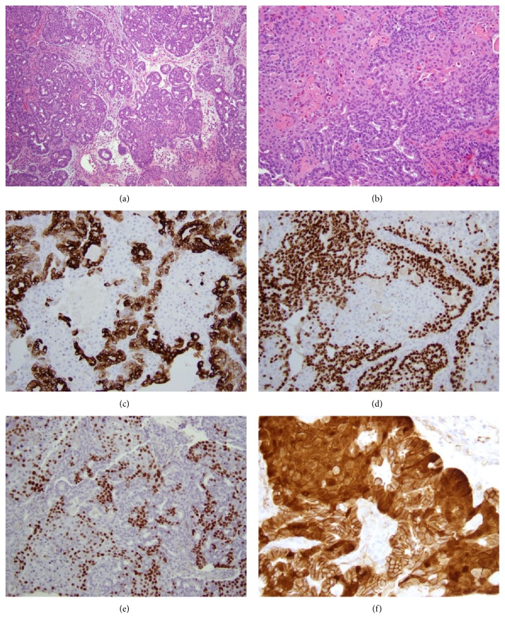
Figure 4.
Morphologic and immunohistochemical findings in Case 2. The tumor is composed of a tubulolobular proliferation of cuboidal cells with minimal cytologic atypia, and interspersed areas of squamoid morular metaplasia ((a), H&E, x100). Other areas of the tumor show more confluent squamoid metaplasia ((b), H&E, x200). CK7 positivity is restricted to glandular areas of the tumor ((c), x200). SOX10 is likewise positive in glandular areas and negative in squamoid areas ((d), x200). CDX2 shows an inverse staining pattern to CK7 and SOX10, with positivity restricted to squamoid areas ((e), x200). Nuclear accumulation of β catenin is seen in squamoid areas ((f), x400).

