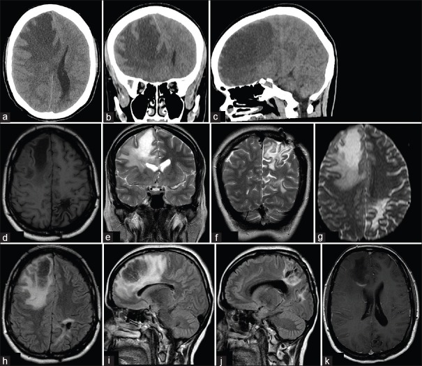
Figure 1.
Neuroimage is showing tumefactive demyelinating area. Axial (a), coronal (b), and sagittal (c) view of noncontrast head computed tomography scan showing a hypodense area in the right frontal lobe, with mass effect and subfalcine herniation. Axial T1-weighted (d), Coronal T2-weighted (e and f), Axial diffusion-weighted (g), Axial (h), and sagittal (i and j) Fluid-attenuated inversion recovery. Axial contrast showing “C”-shaped ring enhancement (k). The magnetic resonance imaging images show the left parietal lobe where there is a residual lesion from the first attack (d, f, g-j); and the right frontal lobe, with the second new lesion (d, e, g-i, k)

