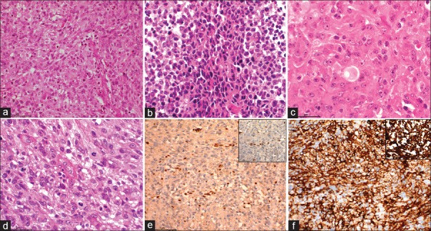
Figure 4.
(a) Microphotograph showing a densely cellular neoplasm (H and E, ×100). (b) Microphotograph showing small/primitive looking cells arranged in sheets (H and E, ×200). (c) Microphotograph showing predominantly rhabdoid cells (H and E, ×400). (d) Microphotograph showing neoplastic cells exhibiting marked pleomorphism and aberrant mitosis (H and E, ×400). (e) Microphotograph showing loss of nuclear staining of integrase interactor 1 with positively stained endothelial cells acting as internal control for the stain (integrase interactor 1 IHC, ×200, inset ×400). (f) Microphotograph showing diffuse and variable positivity for epithelial membrane antigen (IHC for epithelial membrane antigen × 200) and inset showing strong positive staining for vimentin (IHC for vimentin ×400)

