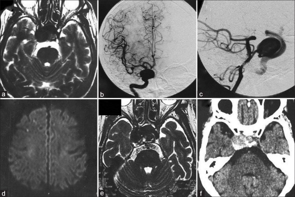
Figure 1.
T2-weighted magnetic resonance image showing a large intracavernous internal carotid artery aneurysm (a), and cerebral digital subtraction angiograms revealing a large aneurysm at the internal carotid artery-primitive trigeminal artery bifurcation (b and c), with maximum diameter of 18 mm. Three years later, diffusion-weighted magnetic resonance image showing scattered cerebral infarctions in the right hemisphere (d), and T2-weighted magnetic resonance image demonstrating enlargement and partial thrombosis of the aneurysm, with maximum diameter of 22 mm (e). Computed tomography scan showing partial thrombosis in the aneurysm sac (f)

