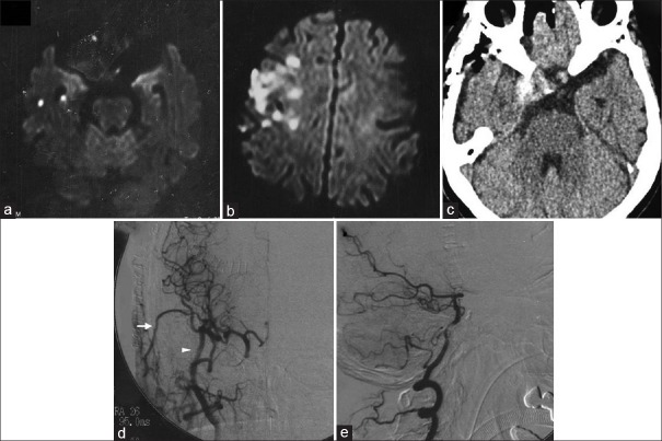
Figure 2.
Diffusion-weighted magnetic resonance image showing new infarctions in the right temporal lobe (a). Six days later, diffusion-weighted magnetic resonance image revealing recurrence of infarction in the right hemisphere (b). Computed tomography scan on the day after the operation showing thrombosis of the aneurysm (c). Cerebral digital subtraction angiogram of the right carotid artery 5 days after the operation demonstrating good patency of bypasses (arrow: superficial temporal artery, arrowhead: radial artery graft) and disappearance of anterograde flow to the aneurysm (d). Digital subtraction angiogram of the right vertebral artery also demonstrating disappearance of anterograde flow to the aneurysm through the primitive trigeminal artery (e)

