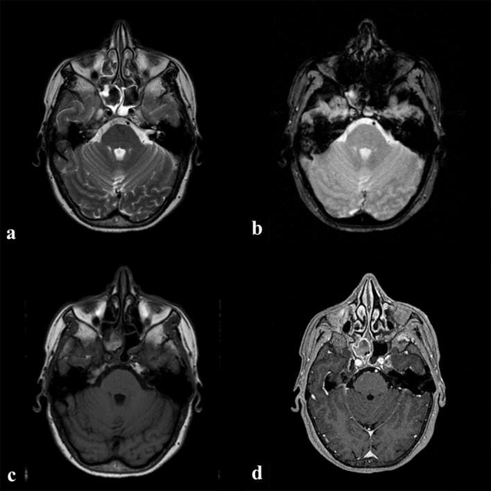
Figure 1.
MRI examination: the right sphenoid sinus is filled with a soft tissue mass that appears hypointense on T2 weighted image (a) with some susceptibility blooming artefacts on T2* images (b) and is hyperintense on T1 weighted image; (c) on post-contrast MPRAGE T1 images (d) the mass did not enhance, whereas the inflammatory mucosa underwent Contrast Enhancement (CE).

