Figure 8.

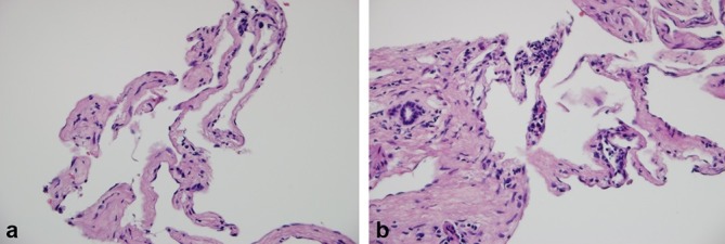
Liver biopsy. (a) Vascular channels lined by endothelial cells without dysplasia, separated by septa of fibrous enlargement (100×); (b) Vascular channels adjacent to a portal space that presents occasional lymphocytes and fibrosis (200×).

Liver biopsy. (a) Vascular channels lined by endothelial cells without dysplasia, separated by septa of fibrous enlargement (100×); (b) Vascular channels adjacent to a portal space that presents occasional lymphocytes and fibrosis (200×).