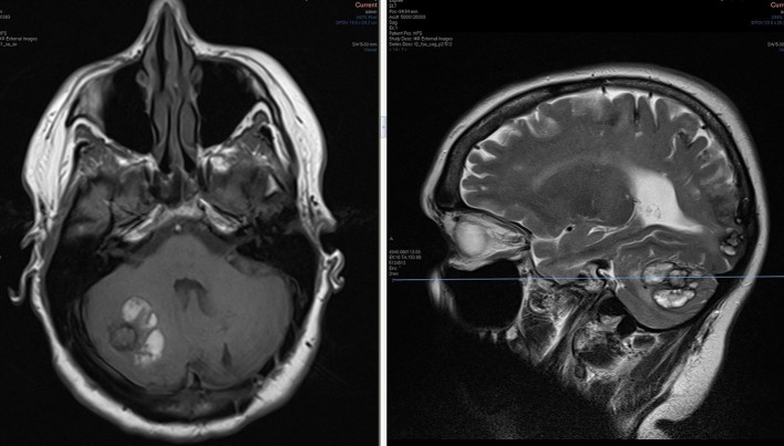
Figure 3.
Non-contrast T1 (left) and T2 (right) images at the level of an irregular ring- like lesion in the right cerebellar hemisphere. The lesion demonstrates low T1 signal and high T2 signal, compatible with a metastasis. Classically a melanoma deposit would demonstrate high T1 signal. Hyperintense T1 and T2 material surrounding the lesion is in keeping with subacute haemorrhage. Note the additional site of haemorrhage in the inferior right occipital lobe, seen on the sagittal slice.

