Figure 1.

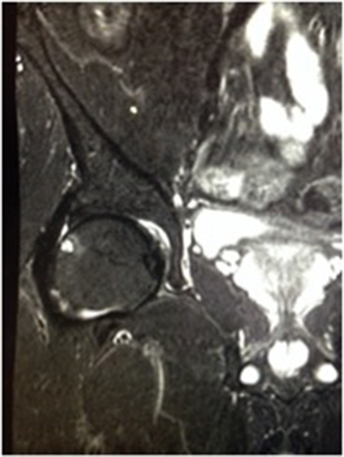
Coronal short tau inversion-recovery image of the right hip demonstrating a 5-mm herniation pit at the anterior aspect of the superolateral femoral head–neck junction with mild surrounding bone oedema.

Coronal short tau inversion-recovery image of the right hip demonstrating a 5-mm herniation pit at the anterior aspect of the superolateral femoral head–neck junction with mild surrounding bone oedema.