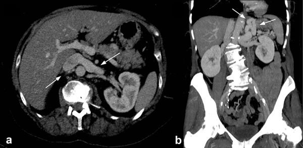
Figure 1.

(a) Axial CT image (1 mm, maximum intensity projection) showing an engorged left renal vein posteriorly, relative to the portal vein, due to the congenital extrahepatic portosystemic shunt in a 68-year-old asymptomatic female patient. (b) Coronal reconstructed CT image (5 mm, maximum intensity projection) showing a tortuous and dilated left gastric (shunt) and splenic vein within the left upper abdomen and communicating superior mesenteric vein and portal vein with the left renal vein and inferior vena cava (Supplementary Video 1a,b).
