
Figure 5.
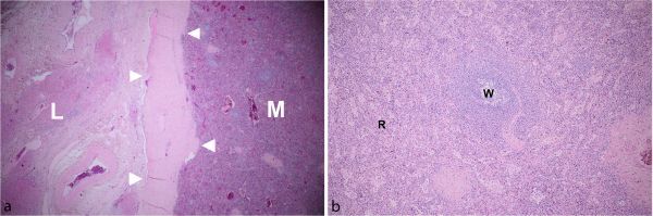
Microscopic examination (low power field, 4×) shows the interface between a well-encapsulated mass with thick fibrous capsule and adjacent liver parenchyma (a), containing red and white pulp (b). (L, liver; M, mass; R, red pulp; W, white pulp; arrowheads, fibrous capsule).