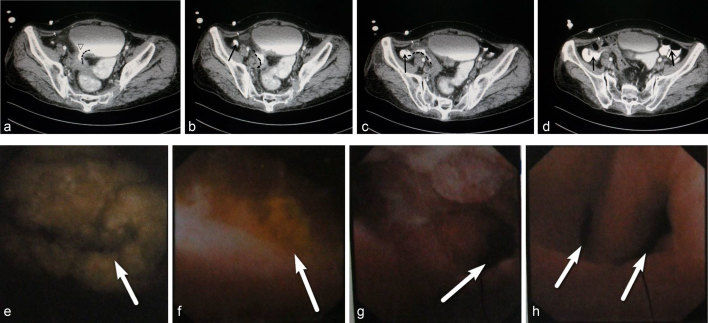
Figure 2.
(a, b) CT scan revealed the right aspect of the bladder wall to be thickened (maximum thickness, ~0.97 cm). The fistula (dotted curved line) was visible. On the right posterior wall of the bladder, a discontinuous region of ~1 cm was noted (triangle). (c, d) Contrast agent was observed in the bladder, and part of the intestine (which appeared to be the ileum) was conjoined. Contrast agent filled the caecum (arrowhead). (e) Cystoscopy demonstrated a mass in the bladder with white stones (arrow) within it (size, 2.5 × 1.5 cm). (f) On the right aspect of the wall was a fistula (diameter, ~1 cm) that was associated with pus and yellow floccules (arrow). (g) Opening of the fistula (arrow). (h) The fistula (arrow) bifurcates at ≈2 cm.

