Figure 1.

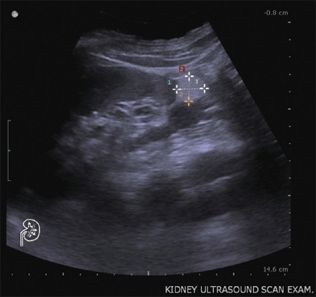
Kidney ultrasound. Ultrasound reveals a homogeneous, well-defined, hyperechoic lesion in the left lower kidney, which demonstrates the presence of macroscopic fat. Renal angiomyolipoma is the most likely diagnosis

Kidney ultrasound. Ultrasound reveals a homogeneous, well-defined, hyperechoic lesion in the left lower kidney, which demonstrates the presence of macroscopic fat. Renal angiomyolipoma is the most likely diagnosis