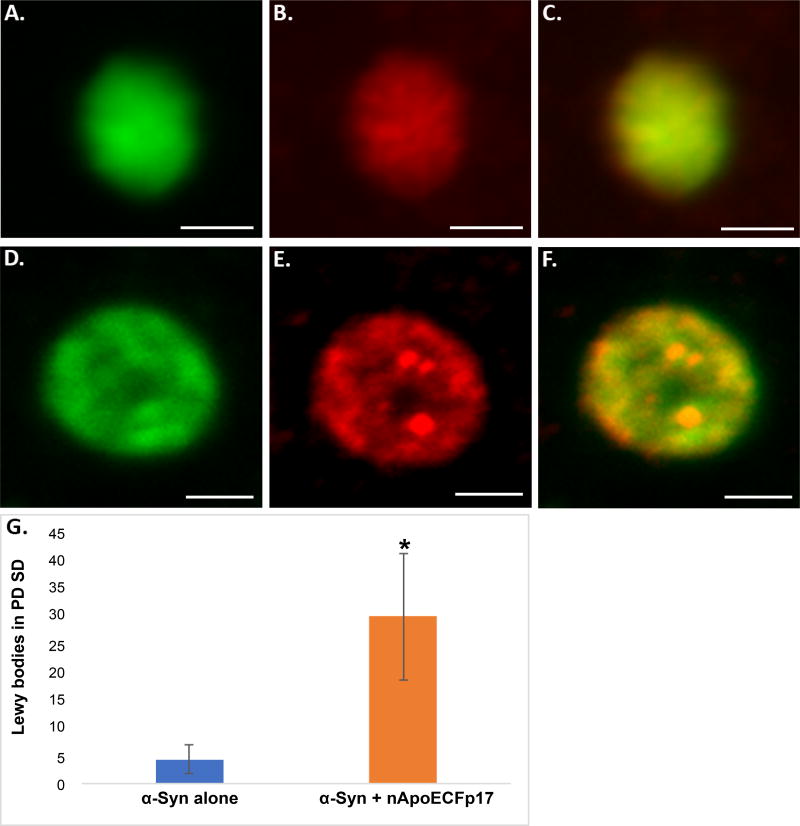
Figure 2. Localization of an amino-terminal fragment of apoE within Lewy bodies of the human PD brain (A–F).
Representation images from confocal immunofluorescence in two different PD cases utilizing antibodies nApoECFp17 (A and D), α-Syn (B and E), with the merged images shown in (C and F). Strong co-localization of the two antibodies was observed in Lewy bodies of the PD brain; (G): Quantification of the number of Lewy bodies doubled-labeled with nApoECFp17 and α-Syn indicated co-localization in 87.5% of the total number of Lewy bodies identified in the substantia nigra. Data depict the number of α-Syn-labeled Lewy bodies alone (blue bar) and the number of Lewy bodies labeled with both α-Syn and nApoECFp17 (orange bar) identified in substantia nigra PD sections by immunofluorescence microscopy (n = 3 different PD cases ± S.D.). All scale bars represent 5 µm. Asterisk denotes significant difference, p = 0.018.

