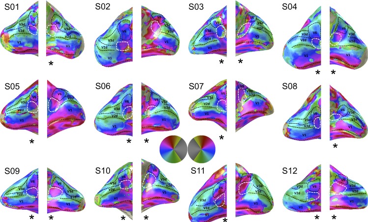
Figure 1.
Wide-field retinotopic mapping results for all twelve subjects. Shown are polar angle maps. The borders between V1, V2, and V3 are indicated by the dashed black lines. V2A is enclosed by a dashed white line and can be found inferior to area V6 at the anterior end of V2d. V6 is enclosed by the dashed black line. The asterisks beneath the hemispheres indicate if we can identify the hemifield representation after careful inspection of the figure (17 of the 24 hemispheres in 11 of the 12 participants). Note in four of the other seven hemispheres we can identify an upper field representation only (S2 left hemisphere, S5 right hemisphere, S8 left hemisphere, and S11 right hemisphere).

