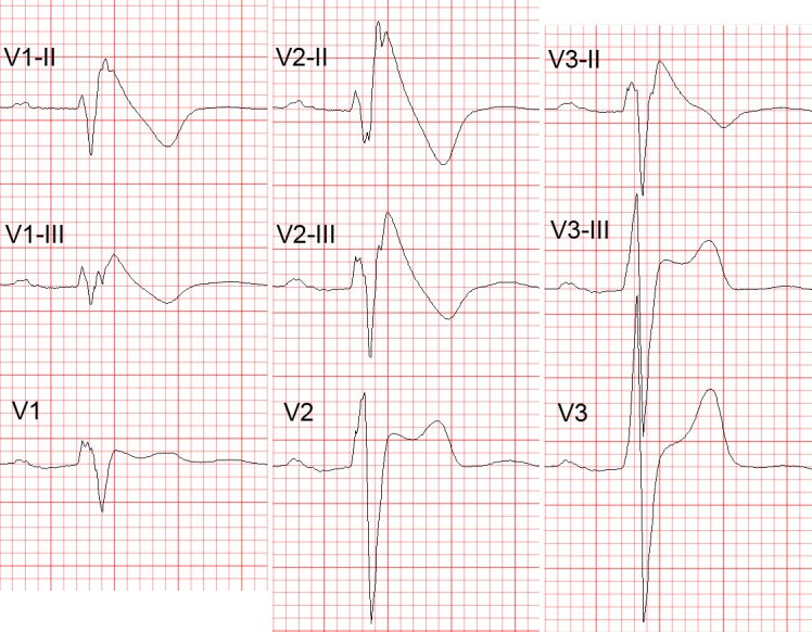
Figure 2: Diagnostic Value of Higher Intercostal Positions.

Recording Leads V1 to V3 from One or Two Intercostal (i.c.) spaces higher than their standard positions (i.e. 3rd or 2nd i.c. space for leads V1 and V2) increases their sensitivity for detecting the diagnostic type 1 Brugada ECG pattern. Note the transition of the ECG complexes in lead V3 from a normal morphology (in the standard position) to type 2 (one i.c. space higher) and type 1 Brugada ECG pattern (two i.c. spaces higher). V1-III, V2-III, V1-II, V2-II = leads V1 and V2 from the 3rd and 2nd i.c. spaces, respectively; V3-III, V3-II = lead V3 recorded one and two spaces higher, respectively. ECG = electrocardiogram.
