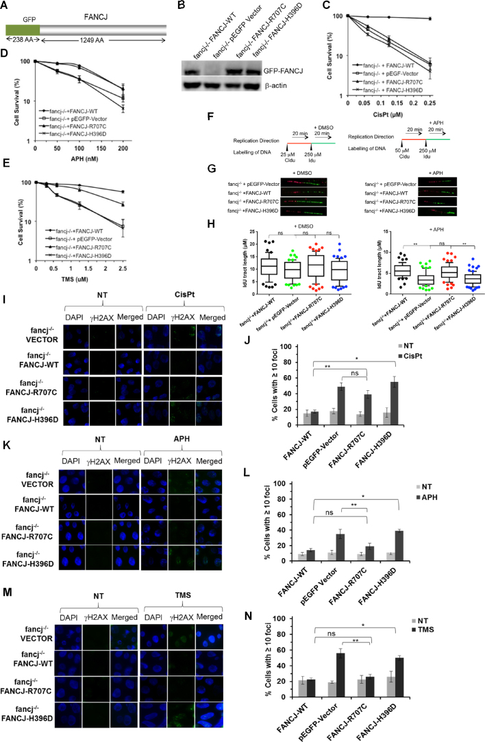
Figure 5.
FANCJ-R707C is a separation-of-function mutant that partially rescues sensitivity to the replication stress inducing agents aphidicolin or telomestatin but fails to restore cisplatin resistance. (A) Schematic diagram of green fluorescence protein (GFP)-FANCJ recombinant protein expressed in fancj−/− cells for genetic complementation assays. (B) Western blot analysis of whole cell lysate protein (40 μg) from fancj−/− DT40 cells transfected with plasmid encoding (pEGFP-Vector), pEGFP-FANCJ-WT (FANCJ-WT), pEGFP-FANCJ-R707C (FANCJ-R707C), or pEGFP-FANCJ-H396D (FANCJ-H396D). Protein was detected with antibody against FANCJ or actin (as a loading control, 10% loaded). (C-E) Cell survival assays, as measured by methyl cellulose colony formation, for the indicated fancj−/− cells expressing mutant or wild-type FANCJ proteins. Cells were exposed to the indicated concentrations of CisPt (C), APH (D) or TMS (E). Filled square, fancj−/− cells transfected with FANCJ-WT; open square, fancj−/− cells transfected with pEGFP-Vector; filled triangle, fancj−/− cells transfected with FANCJ-R707C; open cross- fancj−/− cells transfected with FANCJ-H396D. (F, G) FANCJ-R707C restores normal replication restart after APH exposure. Schematic representation of the protocol used to track DNA replication fibers is shown in Panel F. Cells were pulse-chased with CldU (red label), and then labeled with IdU (green label) for the indicated times. (G) Representative images of fluorescently-labeled DNA fibers from the indicated cell lines treated with DMSO or APH. (H) Box and whiskers graphs indicating the 10–90 percentile of the IdU tract length (μm) for ongoing forks. The data, presented as mean ± standard error of the mean (s.e.m.), are based on the measurement of at least 100 DNA fibers from two independent experiments. (I–N), DNA damage, as marked by immunofluorescent detection of γ-H2AX foci, in the indicated fancj−/− transfected cell lines. fancj−/− cells transfected with pEGFP-Vector, pEGFP-FANCJ-WT (FANCJ-WT), pEGFP-FANCJ-R707C (FANCJ-R707C) or pEGFP-FANCJ-H396D (FANCJ-H396D) were exposed for 14 h to: CisPt (1 μM) (I, J); APH (200 nM) (K, L); TMS (5 μM) (M, N). Immunofluorescence detection of γ-H2AX foci by Alexa fluor 488 is shown along with DAPI, or merged (DAPI and Alexa fluor 488). Quantitative analyses of γ-H2AX foci are shown with S.D. (ns-not significant; **P < 0.05, *P < 0.01 (Student's t-test).

