Figure 5.
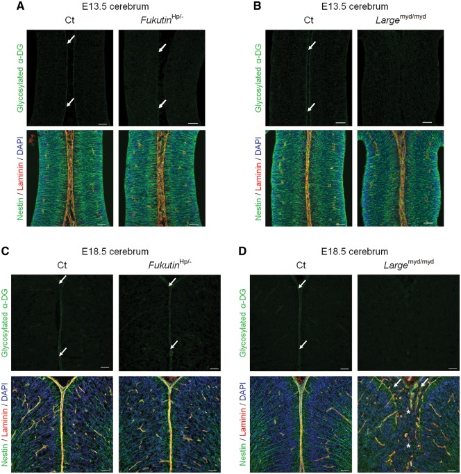
Dystroglycan glycosylation and phenotypic correlation during brain development in other mouse models of dystroglycanopathy. (A, B) Immunofluorescence analysis of the developing cortex at E13.5. (A, upper panel) FukutinHp/- mice demonstrated residual α-DG glycosylation at the glia limitans (arrow). (B, upper panel) α-DG glycosylation was defective in Largemyd/myd mice. (A and B, lower panel) Both strains exhibited almost normal brain structure regardless of α-DG glycosylation state. (C, D) Immunofluorescence analysis of the developing cortex at E18.5. (C, upper panel) Glycosylation of α-DG was observed at the glia limitans in FukutinHp/- mice (arrow). (D, upper panel) Glycosylation of α-DG was consistently defective in Largemyd/myd mice. (C, lower panel) Almost no structural defects were observed in FukutinHp/- mice. (D, lower panel) Diffuse and severe brain malformations were observed in Largemyd/myd mice. Cerebral hemispheres were widely fused (asterisk), and the glia limitans-basement membrane complex was diffusely dissociated in Largemyd/myd mice, similar to findings observed in Emx1-fukutin-cKO mice. Many ectopic cells were observed in the subarachnoid space (arrow). Scale bars = (A, B, C, D) 50 μm.

