Figure 3.
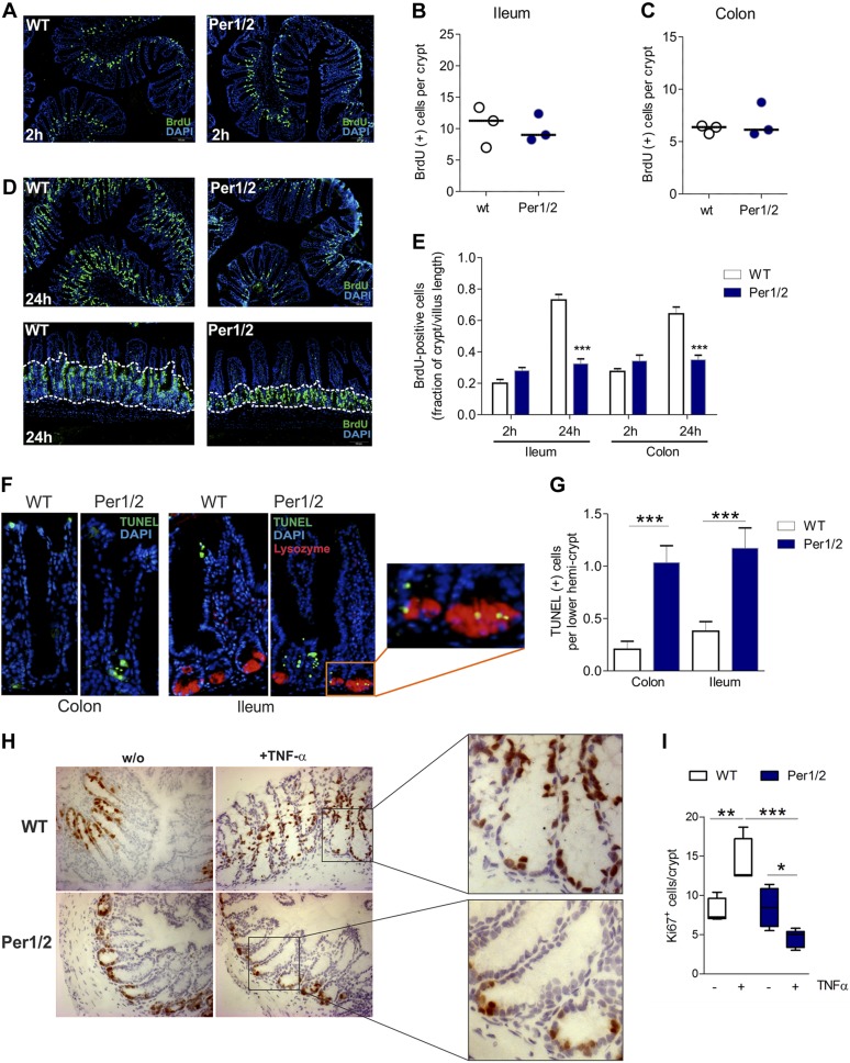
Per1/2 deficiency is accompanied by impaired proliferation and increased cell death in lower hemicrypts. A) Representative images of BrdU-labeled proliferating cells in colonic tissue after 2 h of pulsing. Original magnification, ×10. B, C) Quantification of BrdU+ epithelial cells in ileal (B) and colonic(C) crypts. Mean of 8 distinct tissue slide regions (n = 3 mice/strain). D) Representative images of BrdU-labeled proliferating cells in colonic tissue sections after 24 h of pulsing. Original magnification, ×10 (top); ×20 (bottom). E) Quantification of BrdU+ cells in ileal and colonic tissue specimens after 2 or 24 h of BrdU pulsing. Data are presented as the fraction of BrdU+ cells per crypts in relation to the villus length (n = 3 mice per strain and time point). F) Representative images of TUNEL+ cells in colonic and ileal tissue sections. Inset: colocalization of lysozyme and TUNEL in PCs. G) Quantification of positively labeled cells in colonic and ileal biopsies (n > 8 per strain). H) Representative images of Ki-67-stained cells in colonic tissues in baseline conditions or after TNF-α treatment of WT or Per1/2 mice. Original magnification, ×10, insets: lower crypts. I) Quantification of Ki67+ cells in colonic biopsies (n > 3 per strain). All values are means ± sem. *P < 0.05, **P < 0.01, ***P < 0.001.

