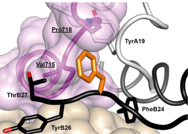
Figure 16.

Atomic environment of PheB24 within the Site 1 μIR complex. Schematic shows all side chains of all residues within 4 Å of that of insulin PheB24 (unlabeled, orange). Insulin A chain is in white, insulin B chain is in black, L1 domain of the insulin receptor is in tan, αCT segment of the insulin receptor is in magenta. Receptor residues have underlined labels whereas insulin residues are in normal font. The schematic is based on PDB entry 4OGA [41].
