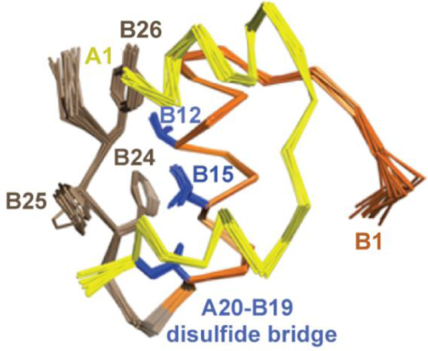
Figure 4.

1H-NMR spectroscopy defines T-like conformation of an engineered insulin monomer. Solution structure of DKP-insulin highlighting the aromatic triplet (PheB24, PheB25 and TyrB26) in brown relative to ValB12, LeuB15 and cysteine B19-A20 in blue. In this monomeric analog dimerization was impaired by the paired substitutions LysB28 and ProB29 [57], and the trimer interface was disrupted by AspB10 [58, 59]. The A chain is shown in yellow, and B chain in orange (B1-B19) and brown (B20-B30).
