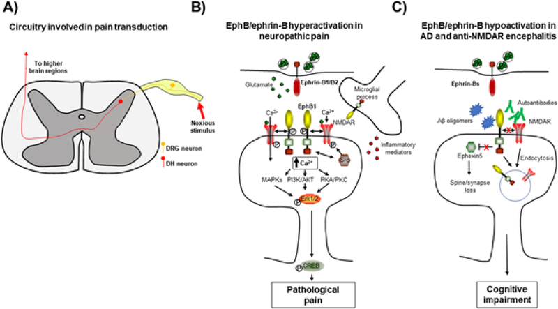
Figure 4.
Eph-ephrin signaling in disease. (A) Model of spinal circuitry involved in pain transduction. (B) Pathological increases in EphB1 signaling in dorsal horn neurons result in neuropathic pain. Activation of EphB1 by pre-synaptic ephrin-Bs results in enhanced NMDAR currents, activating MAPK, PI3K/AKT, PKA and PKC signaling pathways and inducing pathological pain. (C) Disruption of post-synaptic EphB signaling in Alzheimer’s disease (AD) and anti-NMDAR encephalitis results in cognitive impairment. In AD, Aβ oligomers bind to EphB2, which results in depletion of synaptic NMDARs either through disruption of the EphB2-NMDAR interaction or by an as yet undetermined mechanism involving the PDZ binding domain of EphB2. Knockout of Ephexin5 rescues behavioral deficits in APP mice, suggesting that reduced levels of EphB2, which normally inhibits Ephexin5, may play a role in synapse loss in this mouse model of AD. In anti-NMDAR encephalitis, autoantibodies bind to NMDARs and disrupt the EphB2-NMDAR interaction, resulting in endocytosis of NMDARs.

