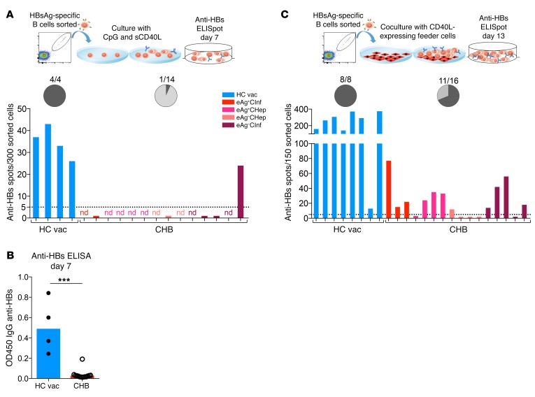
Figure 4. HBsAg-specific B cells from CHB patients are dysfunctional and require coculture with CD40L-expressing feeder cells for survival, expansion, and anti-HBs production.
(A) HBsAg-specific B cells from 4 healthy vaccinated donors and 14 CHB patients were FACS sorted and cultured in the presence of CpG, sCD40L, IL-2, IL-10, and IL-15 for 4 days and subsequently with IL-2, IL-6, IL-10, and IL-15 for another 3 days before the anti-HBs ELISpot assays were performed. nd, not done due to low cell number. The chart shows the percentage of anti-HBs producers. Dark gray indicates positive. (B) Supernatants of the cultured specific B cells (from A) were taken after 7 days culture, and anti-HBs levels were measured by ELISA. Data are presented as median, and statistical analysis was performed by Mann-Whitney U test. ***P < 0.001. (C) HBsAg-specific B cells from 8 healthy vaccinated donors and 16 CHB patients were FACS sorted and cocultured with CD40L-expressing feeder cells in the presence of IL-2 and IL-21 for 13 days before anti-HBs ELISpot assays were performed. Chart shows the percentage of anti-HBs producers. Dark gray indicates positive.

