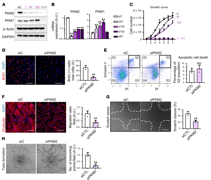
Figure 2. PKM2 is crucial for EC proliferation and migration in vitro.
(A) Western blot analysis of PKM2 and PKM1 protein expression in HUVECs demonstrates that the siRNAs used in this study give PKM2-specific and efficient knockdown. qPCR analysis of PKM2 and PKM1 mRNA expression. Note that the y axis for PKM2 is a log scale (n = 3). (C) Growth curve of HUVECs with various siRNAs targeting PKM2. The efficiency of knockdown shown in B correlates with the effect on proliferation (n = 5). (D) Percentage of BrdU+ cells is reduced in siPKM2 ECs (n = 3). (E) Apoptotic cell death, assessed by Annexin V and PI staining, is not induced in siPKM2 ECs (n = 3). (F) Reduction in transwell migration of ECs by siPKM2. Cells that migrated across the transwell membrane were visualized by staining with phalloidin (red) and DAPI (blue) (n = 3). (G) Scratch closure was retarded in ECs with siPKM2 (n = 3). (H) Tube formation on Matrigel was impaired in ECs with siPKM2 (n = 3). Scale bars, 100 μm. All data are mean ± SD. **P < 0.01, by 2-tailed Student’s t test.

