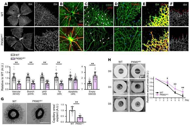
Figure 3. PKM2 is crucial for angiogenesis in vivo.
(A–F) Staining of WT versus PKM2ΔEC mouse retina at P7. (A) The entire retinal vasculature is visualized by IB4 staining. Scale bar, 1 mm. (B) Arterio-venous patterning is analyzed by IB4 and SMA staining. a, artery; v, vein. (C) The number of pH3+ ECs (white arrows) is analyzed by costaining of pH3 and CD31. (D) Apoptosis was analyzed by TUNEL staining. (E) Vascular regression is assessed by empty sleeves (blue arrows) with IB4-negative and collagen IV–positive staining. (F) Number of tip cells (red arrows) is visualized via IB4 staining. Quantifications are at bottom of figure. Scale bars, 50 μm (B–F). Data are mean ± SD of 4–8 different images per mouse (n = 8). **P < 0.01, by 2-tailed Student’s t test. (G) Capillary sprouting of aortic explants from WT versus PKM2ΔEC mouse. At day 3 of aortic ring explant incubation, each explant was photographed and the area of capillary outgrowth was quantified using Image J. Scale bar, 1 mm. Data are mean ± SD; n = 6 segments per aorta from 3 mice each group. **P < 0.01, by 2-tailed Student’s t test. (H) Wound healing assay in WT versus PKM2ΔEC mouse. Two wounds (5 mm) were made on the back of each mouse by using biopsy punch and wound size was monitored every 2–3 days for a week. Data are mean ± SD (n = 5). *P < 0.05, **P < 0.01, by 2-tailed Student’s t test.

