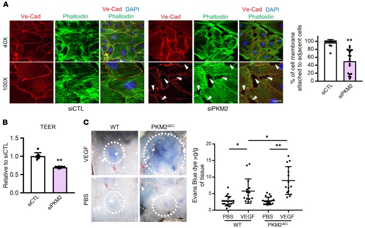
Figure 7. PKM2 is required for vascular barrier function in confluent contact-inhibited ECs.
(A) Disrupted formation of VE-cadherin (VE-Cad) adherent junction with siPKM2. Two days after siRNA transfection, HUVECs were trypsinized and same number of cells was reseeded and allowed to form junctions. At 24 hours after reseeding, cells were immunostained with VE-Cad (red), phalloidin (green), and DAPI (blue). Confocal analysis demonstrates continuous VE-Cad distribution around the entire periphery of the cells with siCTL whereas siPKM2 cells show discontinuous and unstable junctions with intercellular gaps (white arrows). Quantification of cell membrane (%) attached to adjacent cells in siCTL versus siPKM2 HUVECs. siPKM2 cells have less attachment to adjacent cells compared with siCTL (n = 6). Scale bar, 10 μm. (B) Changes in trans-endothelial electrical resistance (TEER) was measured in siCTL versus siPKM2 HUVECs on an electric cell-substrate impedance sensor (ECIS) in 8W1E+ plate at 4,000 Hz (n = 8). (C) Acute vascular hyperpermeability was assessed in WT versus PKM2ΔEC mouse via miles assay. Mice were injected intravenously with Evans Blue dye and were subsequently injected intradermally with PBS and VEGF (100 ng). Representative skin images in response to PBS or VEGF intradermal injections. Scale bar, 50 mm. Quantification of extracted Evans Blue dye normalized to tissue weight (μg of Evans Blue dye / g of tissue; n = 15 each group). All data are mean ± SD. *P < 0.05, **P < 0.01, by 2-tailed Student’s t test.

