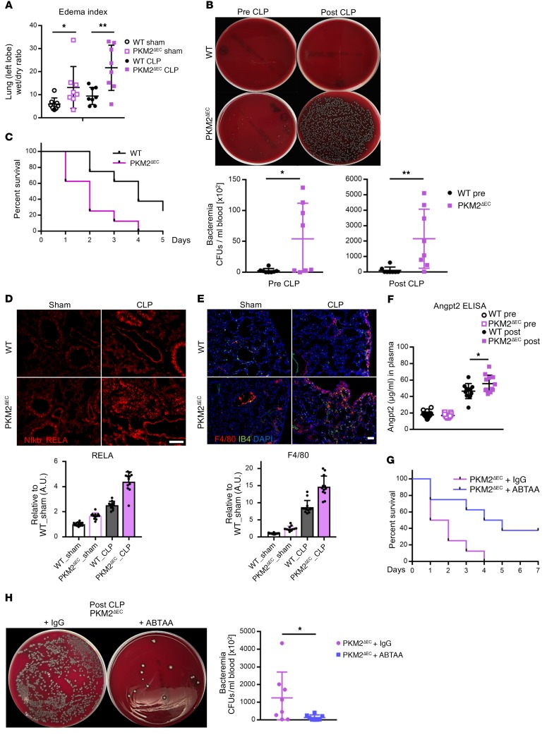
Figure 9. Deletion of endothelial PKM2 exacerbates sepsis-induced responses and mortality in CLP model.
(A) Lung edema was measured as the ratio of the wet weight to the dry weight of the left lobe 20 hours after sham or CLP surgery. Each group used 12- to 14-week-old mice (n = 8 each group). (B) PKM2ΔEC mice have higher bacteremia before and after CLP. Each group used 20- to 22-week-old mice. Representative images of bacterial growth on blood agar and quantification of number of bacterial CFUs in blood (n = 8 each group) are shown. (C) Survival rates of WT and PKM2ΔEC mice after CLP surgery. Each group used 20- to 22-week-old mice (n = 8 each group). (D) Representative immunohistochemistry images of NF-kB RELA subunit (red) 20 hours after sham or CLP surgery. Mice 12- to 14-weeks-old were used. Scale bar, 100 μm. (E) Representative immunohistochemistry images of macrophages (F4/80 in red), endothelial cells (IB4 in green), and nucleus (DAPI in blue). Each group used 12- to 14-week-old mice (n = 12 each group). Scale bar, 50 μm. (F) Plasma levels of ANGPT2 before and after CLP as determined by ELISA analysis. Each group used 12- to 14-week-old mice (n = 12 each group). (G) Survival rates of PKM2ΔEC mice with IgG or ABTAA injection after CLP. Each group used 8- to 10-week-old mice (n = 8 each group). (H) Rescued bacteremia by ABTAA administration in PKM2ΔEC mice after CLP. Representative images of bacterial growth on blood agar. Quantification of number of bacterial CFUs in blood. Each group used 8- to 10-week-old mice (n = 8 each group). All data are mean ± SD. *P < 0.05, **P < 0.01, by 2-tailed Student’s t test.

