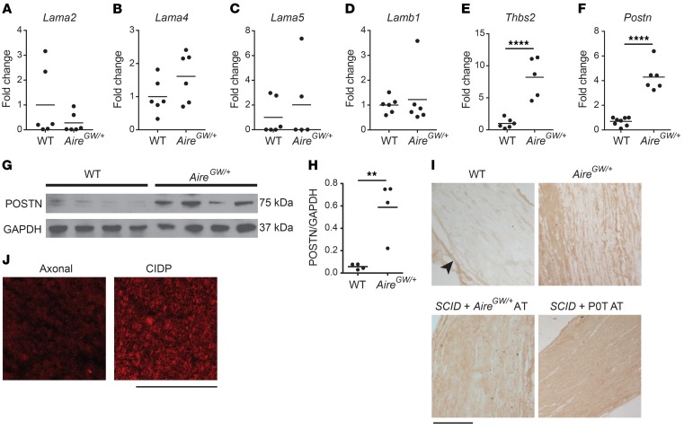
Figure 1. Increased Postn expression in SAPP.
(A–F) RNA was isolated from the sciatic nerves of NOD.WT (WT) and NOD.AireGW/+ neuropathic mice. Lama2 (A), Lama4 (B), Lama5 (C), Lamb1 (D), Thbs2 (E), and Postn (F) expression relative to cyclophilin was measured by qRT-PCR. Values are expressed as the fold change compared with WT. (G) POSTN expression was measured by Western blotting of sciatic nerve lysates from NOD.WT (WT) and NOD.AireGW/+ (AireGW/+) neuropathic mice. GAPDH was used as a loading control. Each lane represents an individual mouse. (H) Densitometric analysis of the Western blot in G. (I) POSTN immunohistochemical staining of sciatic nerves from WT mice, neuropathic NOD.AireGW/+ mice, neuropathic SCID recipients of NOD.AireGW/+ splenocytes, and neuropathic SCID recipients of P0T splenocytes. Note that POSTN immunoreactivity was mostly found in the perineurium (arrowheads) of NOD.WT (WT) nerves, whereas the endoneurium was diffusively positive in nerves from NOD.AireGW/+ mice, SCID recipients of NOD.AireGW/+ splenocyte AT, and SCID recipients of NOD.POT splenocyte AT. Scale bar: 180 μm. (J) Immunofluorescence staining of biopsy samples from patients with axonal neuropathy or CIDP. Increased endoneurial POSTN immunoreactivity was observed in the CIDP sample. Scale bar: 200 μm. (A–F and H) Each dot represents an individual animal. **P < 0.005 and ****P < 0.0001, by 2-tailed, unpaired t test. Individuals values and means are shown.

