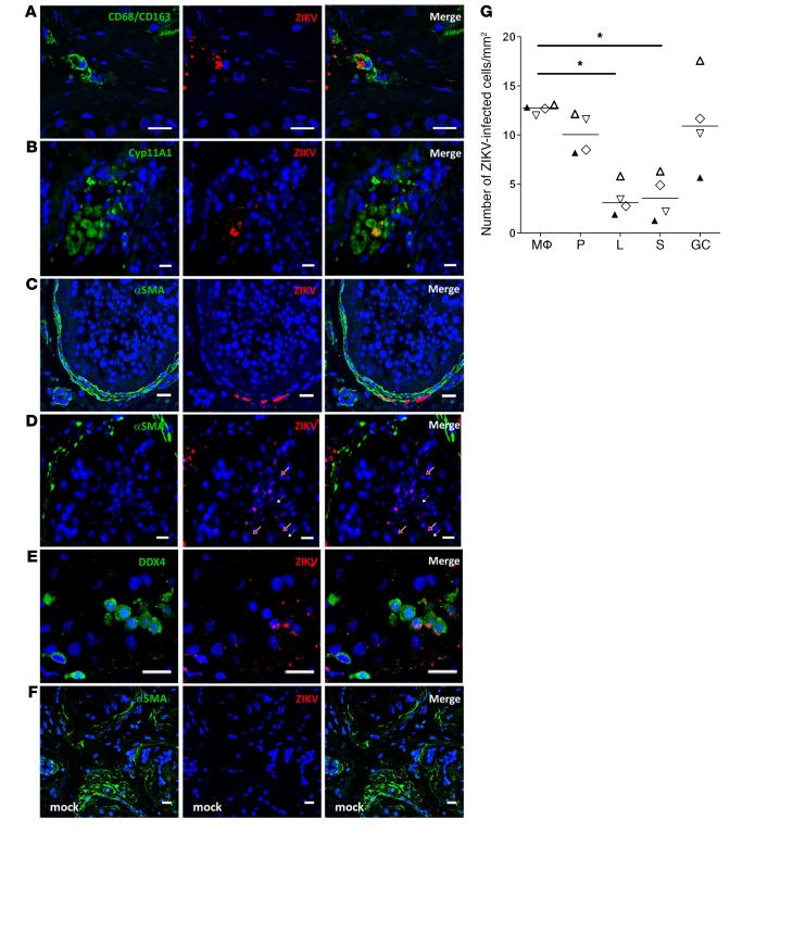
Figure 3. Characterization and quantification of ZIKV-infected human testicular cells ex vivo.
RNAscope ISH for vRNA coupled with immunofluorescence for cell markers identified ZIKV RNA in CD68/CD163+ macrophages (A), Cyp11A1+ Leydig cells (B), α-SMA+ peritubular cells (C), late germ cells localized near the lumen in seminiferous tubules (white arrows, round spermatids; red arrows, elongated spermatids) (D), and DDX4+ early germ cells (E). Staining for ZIKV was not observed in mock-infected testis (F). Nuclei are stained in blue. Scale bars: 20 μm. (G) Infected cells were quantified in at least 3 whole tissue sections from 4 testis donors (each represented by a different symbol) on day 9 p.i. Mϕ, macrophages; P, peritubular cells; L, Leydig cells; S, Sertoli cells; GC: germ cells. Bars represent median. *P < 0.05 (Friedman-Dunn nonparametric comparison).

