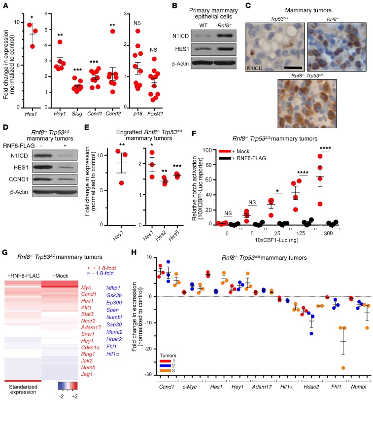
Figure 5. RNF8 negatively regulates Notch signaling in murine mammary luminal progenitors and mammary tumors.
(A) Quantitative reverse transcriptase PCR (RT-qPCR) showing the expression changes of Notch targets and other genes in luminal progenitors from mammary glands of Rnf8–/– females at estrus phase compared with WT littermates. (B) Immunoblot analysis of N1ICD and HES1 in the indicated cells. (C) Representative immunohistochemical analysis of N1ICD levels in the indicated mammary adenocarcinomas (n = 3 each; scale bar: 25 μm). (D) Immunoblot showing expression of the indicated proteins in Rnf8–/– Trp53Δ/Δ mammary tumor cells reconstituted with mock or RNF8-FLAG. (E) RT-qPCR showing increased expression of indicated Notch1 targets in mock-reconstituted, compared with RNF8WT-reconstituted, Rnf8–/– Trp53Δ/Δ mammary tumor cells engrafted for 40 days in NSG mice (see Figure 1C). (F) Notch reporter 10xCBF1-Luc showing relative activation of Notch signaling in mock-reconstituted Rnf8–/– Trp53Δ/Δ mammary tumor cells compared with RNF8WT-reconstituted controls. *P < 0.05, ****P < 0.0001, 2-way ANOVA followed by Tukey’s test. (G) Heatmap showing the standardized expression of Notch targets and/or Notch pathway elements in Rnf8–/– Trp53Δ/Δ mammary tumor cells reconstituted with mock or RNF8-FLAG. (H) Notch targets differentially expressed in RNA-Seq analysis in G were validated by RT-qPCR in 3 different Rnf8–/– Trp53Δ/Δ mammary tumor cell lines and their RNF8-reconstituted controls. Data in B and D are representative of at least 3 experiments. A, E, F, and H: At least 3 experiments were performed, and the means ± SEM are shown. A, E, and H: 2-sided Student’s t test, Rnf8–/– Trp53Δ/Δ mammary tumor cells mock-reconstituted compared with RNF8WT-reconstituted controls. *P < 0.05, **P < 0.01, ***P < 0.001. H: All P values were less than 0.05.

