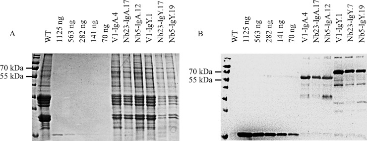
Fig 5. Determination of the concentration of chimeric antibodies present in seed extracts.
Intensities on (A) Coomassie blue stained SDS-PAGE and (B) western blot of protein bands corresponding to a His-tagged nanobody and the ones corresponding to chimeric antibodies were compared. A serial dilution of a His-tagged nanobody with a concentration of 1.2 mg/ml was made. As a negative control, seed extract of wild-type A. thaliana plants was used. The western blot was developed with a mouse anti-histidine tag monoclonal antibody and goat anti-mouse IgG conjugated to HRP.

