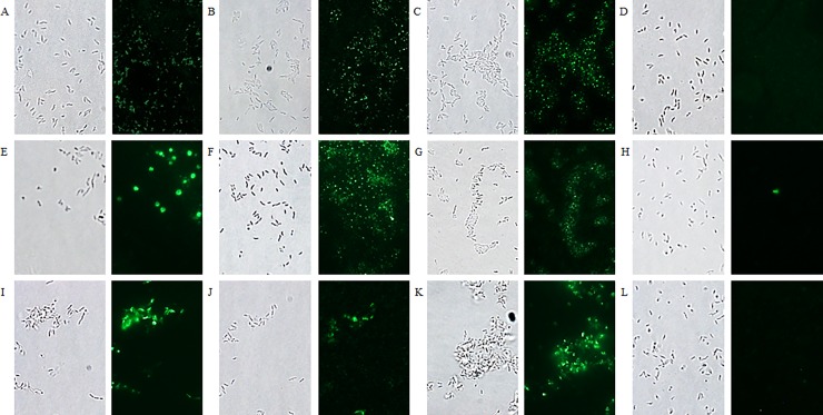
Fig 9. Fluorescence microscopy visualising the binding of labelled anti-flagellin nanobodies with different Campylobacter isolates.
(A, B, C, D) C. jejuni strain KC40, (E, F, G, H) C. jejuni strain Cam12/0156 and (I, J, K, L) C. coli strain K43/5. Interaction with the different isolates is shown with (A, E, I) Nb2Flag8, (B, F, J) Nb2Flag24 and (C, G, K) Nb2Flag67. (D, H, L) The negative control, fluorescently labelled V1 directed against F4-fimbriated E. coli, did not bind with the Campylobacter bacteria. The corresponding bacterial cells were visualised by bright field microscopy.

