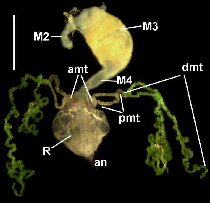
Fig 1. The posterior part of the digestive system of P. apterus with four MTs.

M2, M3, and M4 indicate segments of the midgut; amt–ampullae of MTs; dmt–distal part of MT; pmt–proximal part of MT; an–anus; R–rectum. Reflected light microscopy. Scale bar– 1 mm.
