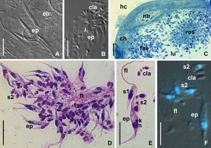
Fig 3. Light microscopy of B. papi in the intestine and MTs.
(A) Epimastigotes in the M3 segment of midgut of diapausing P. apterus (DIC). (B) CLAs in the rectum of ovipositing female (DIC). (C) fragment of transverse semi-thin section of an infected MT (methylene blue staining, BF). (D) rosette-like aggregate (Giemsa-stained smear, BF). (E) Different stages of cyst formation (Giemsa-stained smear, BF). (F) combination of DIC and fluorescence microscopy illustrating the permeation of DAPI into all types of B. papi cells except mature CLAs. ch–cell of the host MT epithelium; cla–cyst-like amastigote; eb–prokaryotic endobionts; ep–epimastigote; fas–fascicule-like aggregate of flagellates; fl–flagellum; hc–hemocoel; k–kinetoplast; lu–lumen of MT; n–nucleus of parasite; nh–nucleus of host cell; ros–rosette-like aggregate of flagellates; s1, s2 –stages of CLAs' formation. Other abbreviations are as in Fig 1. Scale bars: (A–C, E, F)– 10 μm; (D)– 15 μm.

