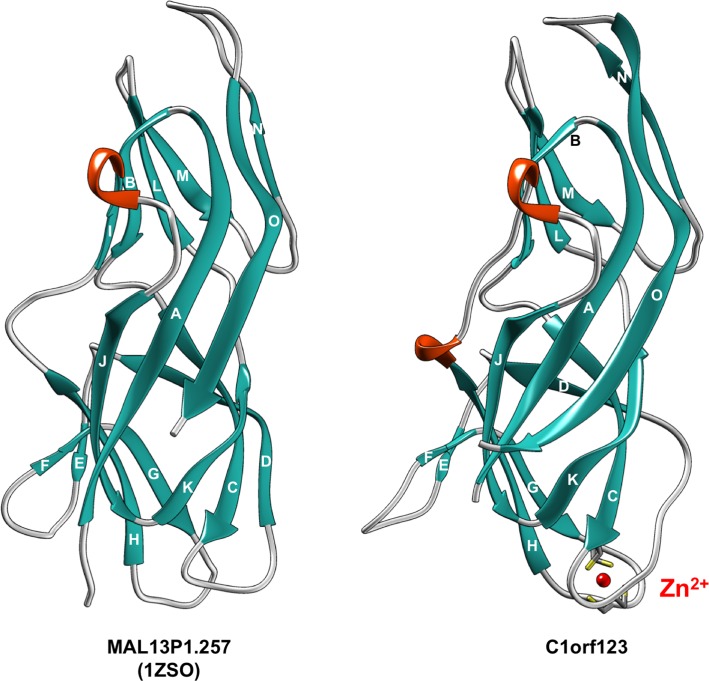
Fig 3. C1orf123 has a similar structure to that of MAL13P1.257.
The structure of C1orf123 revealed in this study is shown as a ribbon model (right) and is compared with the structure of previously published MAL13P1.257 (PDB ID, 1ZSO: left). β-strands are colored in light sea green and designated in alphabetical order from A to O. Helical structures are colored orange. A Zn2+ ion (red ball) ligated with four Cys residues (stick model) is also shown for C1orf123.

