Figure 3.

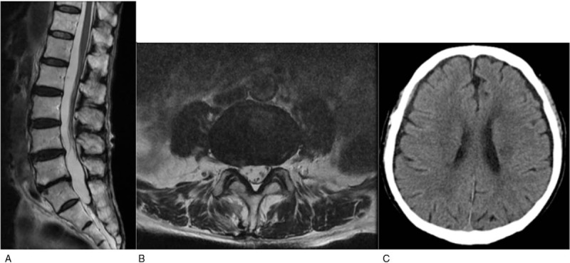
(A, B) Sagittal and axial T2-weighted spine images show complete resorption of the hematoma. (C) Non-contrast computed tomography shows significant resorption of the hematoma.

(A, B) Sagittal and axial T2-weighted spine images show complete resorption of the hematoma. (C) Non-contrast computed tomography shows significant resorption of the hematoma.