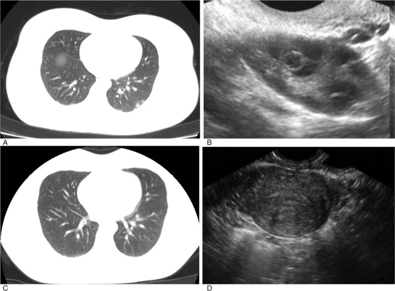
Figure 3.

Radiographic and ultrasound images of the younger sister. (A and B) Before treatment, multiple nodules were displayed in both lungs according to the computed tomography and ultrasonography revealed a 2 cm high echo mass with irregular fluid area inside. (C and D) After 5 cycles of actinomycin D chemotherapy, computed tomography showed most of the primary pulmonary nodules were diminished and the remaining were significantly downsized, while ultrasonography detected no mass in uterine cavity.
