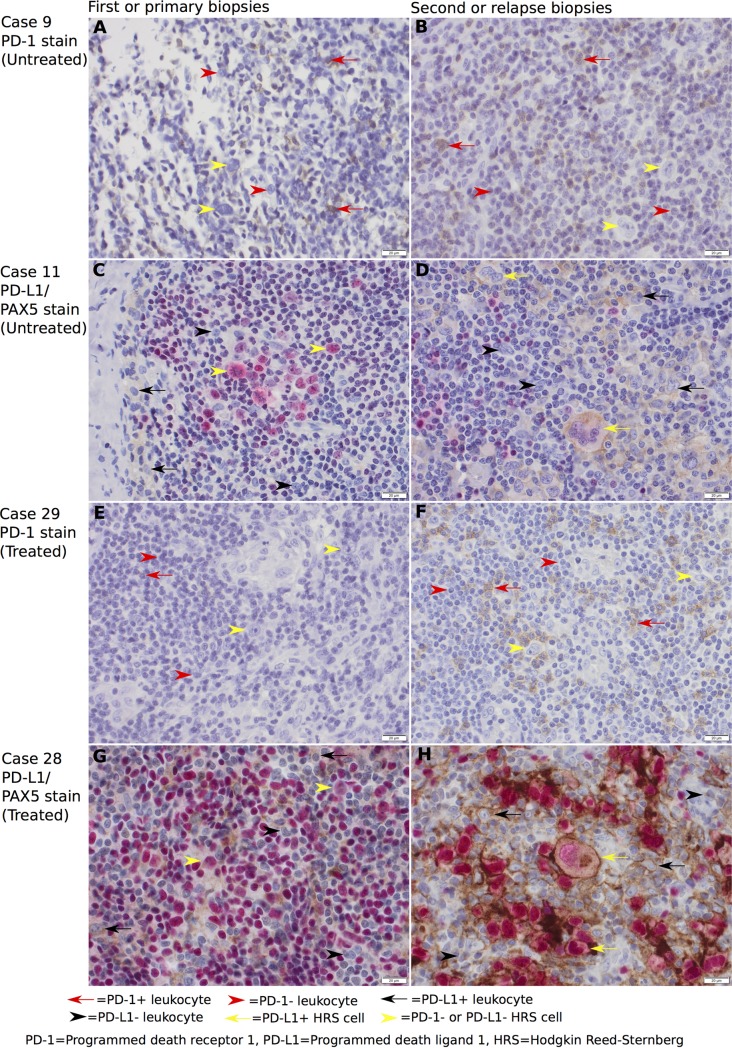
Fig 3.
Representative PD-1 and PD-L1/PAX5 immunohistochemical stainings at 400x magnification with untreated (A-D) and treated (E-H) patients. Brown membranous staining indicates PD-1+ or PD-L1+ cells, while red nuclear staining indicates PAX5+ cells. Untreated: (A) Case 9 biopsy 1 (average 4% PD-1+ leukocytes), (B) case 9 biopsy 2 (average 7% PD-1+ leukocytes), (C) case 11 biopsy 1 (average 5% PD-L1+ leukocytes and 0% PD-L1+ HRS cells), and (D) case 11 biopsy 2 (average 10% PD-L1+ leukocytes and 100% PD-L1+ HRS cells). Treated: (E) Case 29 primary biopsy (average 1% PD-1+ leukocytes), (F) case 29 relapse biopsy (average 16% PD-1+ leukocytes), (G) case 28 primary biopsy (average 10% PD-L1+ leukocytes and 0% PD-L1+ HRS cells), and (H) case 28 relapse biopsy (average 19% PD-L1+ leukocytes and 100% PD-L1+ HRS cells).

