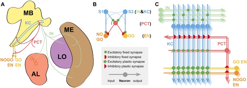
Fig 1. Models of the mushroom bodies based on known neuroanatomy.
A Neuroanatomy: MB Mushroom Bodies; AL Antennal Lobe glomeruli (circles); ME & LO Medulla and Lobula optic neuropils. The relevant neural pathways are shown and labelled for comparison with the model. B Reduced model; neuron classes indicated at righthand side of sub-figure. C Full model, showing the model connectivity and indicating the approximate relative numbers of each neuron type. Colour coding and labels are preserved throughout all the diagrams for clarity. Excitatory and inhibitory connections indicated as in figure legend. Key of neuron types: KC, Kenyon Cells; PCT, Protocerebellar Tract neurons; IN, Input Neurons (olfactory or visual); EN, Extrinsic MB Neurons from the GO and NOGO subpopulations, where the subpopulation with the highest sum activity defines the behavioural choice in the experimental protocol (Fig 4).

