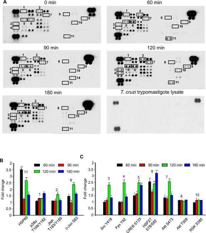
Fig 2. Phosphoproteomic array analysis of T. cruzi-infected human colonic epithelial cells.
Primary HCoEpiC challenged with T. cruzi at multiple time points were lysed and incubated with phosphoproteomic array membranes. (A) Template showing location of kinases and phosphoprotein antibodies spotted onto the human phospho-kinase array membrane. The signals of selected kinases and phosphoproteins at 0, 60, 90, 120, and 180 minutes are indicated by numbers (1–11). Equal amount of T cruzi trypomastigotes lysate was used to evaluate cross reactivity with the membranes. Each signal number was maintained throughout the time course of the experiment. Reference spots are included to align the array membrane and to show that the array has been incubated with streptavidin-HRP. (B) Quantification of mean spot pixel density relative to control represented as fold change for proteins associated with cellular transformation pathways. (C) Quantification of mean spot pixel density relative to control for proteins associated with other cellular pathways. Mean values of biological replicates ± SE are shown. The value of p<0.05 was considered significant. *p<0.05; **p<0.001.

