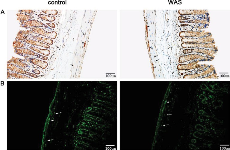
Figure 3.
Immunostaining of CHT1 in the colon.
Notes: (A) Immunohistochemical staining for CHT1 reveals that the AOD of CHT1 in the muscular layer of WAS rats (right) is increased compared to that of control rats (left) (200×). (B) Immunofluorescence staining for CHT1 shows that the density of CHT1 in the muscular layer is significantly elevated after 10 days of WAS (200×). Both the black and white arrows indicate CHT1-positive cells that are mainly fusiform in shape and found within the circular muscle, exhibiting the morphologic characteristics of ICC and smooth muscle cells. Both black and white arrowheads indicate CHT1-positive cells that are predominantly round in shape, located between the circular muscle and longitudinal muscle, showing the hallmark of MP neurons. Bars: (A) = 100 µm, (B) = 100 µm; n of each group = 6.
Abbreviations: AOD, average optical density; ICC, interstitial cells of Cajal; MP, myenteric plexus; WAS, water avoidance stress.

