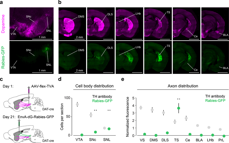
Figure 2. TS-projecting dopamine neurons are mainly localized in lateral substantia nigra and do not send substantial collaterals to other regions.
(a) Midbrain dopamine neurons labelled with anti-TH antibody (magenta) and TS-projecting dopamine neurons labelled with GFP (green). (b) Forebrain dopamine axons (magenta) and the axons of TS-projecting dopamine neurons (green). (c) Schematic for labelling TS-projecting dopamine neurons and their axons. (d) Distribution of GFP-labelled (green) cell bodies and TH-labelled cell bodies in the midbrain (mean ± s.e.m. across n = 6 animals). For SNC: t = 5.042, p = 0.0040, n = 6 animals, t-test. For SNL: t = 9.36, p = 0.00023, n = 6 animals, t-test. (e) Distribution of GFP-labelled axons in the forebrain and distribution of TH-labelled axons (mean ± s.e.m. across n = 8 animals). For TS: t = 7.46, p = 0.00029, n = 8 animals, t-test with Tukey correction for multiple comparisons. ** P < 0.005, two-sided t-test with Tukey correction for multiple comparisons.

