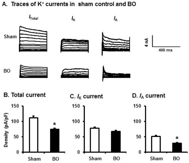
Fig. 4.
Voltage-gated K+ current (Kv) in colon-projecting DRG neurons in sham and BO (day 7) rats. (A) Representative traces of Kv in sham and BO rats, with the Itotal in the left, the IK in the center of the panel, and the IA in the right. For total KV current (Itotal), the membrane potential was held at −100 mV and voltage steps were from −40 to +30 mV with 5-mV increments and 400-ms duration. For sustained Kv current (IK), the membrane potential was held at −50 mV and the voltage steps were the same as above. Currents generated by these two protocols were subtracted to produce IA. Bar graphs of the mean peak Kv density of DRG neurons from sham and BO rats were displayed in B for Itotal, in C for IK, and in D for IA currents. The current density (in pA/pF) was calculated by dividing the current amplitude by cell membrane capacitance. N= 30 neurons (5 rats) in sham and 31 neurons (6 rats) in BO. *p < 0.05 vs. sham.

